Function of the Eye
The eye has many parts that work together to provide the clear vision we view our environment with. Often referred to as a camera, the eye converts light into electrical signals, which when transformed in the brain, give us the images we see of the world.
Structures of the Eye
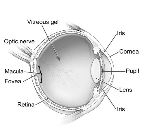
Cornea: The first area that light enters the eye. It is kept moist with constant tear production. Dry eyes can affect the image that passes through this structure, making it blurry.
Pupil: The size of the pupil determines how much light can enter the eye. When it is constricted (small) it allows less light in, and when it is dilated (large), it lets more light in. When your pupil is dilated, it allows for a more thorough internal eye exam.
Iris: This the coloured portion of your eye, and it is responsible for changing the diameter (size) of the pupil in response to the light in your environment.
Lens: This structure focuses the light on your retina, and changes in shape when reading things up close or observing something out in the distance. As we age, it looses its flexibility, limiting the ability to adjust to these changes. This results in the need for reading glasses later in life.
Vitreous: This is the jelly substance that fills the interior of the eye. It is here that is responsible for the production of floaters, small shadows cast in your vision.
Retina: This thin tissue layer acts like the film in the camera, capturing the light and transforming it into the electrical signals for the brain. It is has many blood vessels to nourish it like other organs of the body, and it has areas responsible for fine detail, colour vision, and peripheral vision.
Macula/Fovea: The macula is used for colour vision and the fine details we are able to see. The fovea is at the centre of the macula, where the concentration of the cells needed for central vision is the highest. It is here where diseases such as AMD are so damaging, resulting in a loss of this central vision.
Optic Nerve: the transmission system from the retina, to your brain to produce the images you see in front of you.